
Coronary spasm (CS) is defined as diffuse or focal severe vasoconstriction of the coronary arteries resulting in impaired myocardial perfusion. It was first described in 1959 by Prinzmetal as anginal pain associated with transient ST-segment elevations or depressions on an electrocardiogram (ECG). CS was historically thought to be a condition predominantly affecting middle-aged males in East Asia and was rare in European and North American populations; more recent data suggest that it may be a common cause of myocardial infarction with non-obstructive coronary arteries (MINOCA) in European and North American patients, with a more equal sex distribution. Microvascular spasm has been recognised in the last 20 years and is more poorly characterised. CS, both epicardial and microvascular, remains an important and likely underdiagnosed cause of chronic chest pain in the outpatient setting. We describe advances in our understanding and the definition of CS, techniques for diagnosis, epidemiology and the natural history of the condition, and therapeutic modalities.
Coronary spasm and vasomotor dysfunction
Initially, CS was considered a disease of the epicardial coronary arteries, but, as understanding increased, spasm of the arterioles within the microcirculation was also identified. CS is best considered as part of a spectrum of vasomotor disorders affecting the coronary arteries and may coexist with coronary microvascular dysfunction (CMD). Patients with vasomotor disorders have dysregulation of vasoconstriction and vasodilatation of the coronary vasculature, leading to episodic ischaemia and chest pain. Patients may present with MINOCA or sudden cardiac death or, more commonly, with chronic chest pain – either angina with non-obstructive coronary arteries (ANOCA) or ischaemia with non-obstructive coronary arteries (INOCA).
Pathophysiology
The coronary arteries are dynamic regulators of coronary flow; they constrict and dilate to regulate myocardial perfusion and match supply to demand. Vasodilation occurs due to an increase in endothelium-derived relaxing factors including nitric oxide (NO) and relaxation of the vascular smooth muscle cells (VSMCs). The principal abnormality in CS is hyperreactivity of the VSMCs within the media, with increased sensitivity to intracellular calcium concentrations that results in an increased propensity towards hypercontraction. The endothelium and the perivascular adipose tissue (PVAT) within the adventitia both secrete signalling factors that modulate vasoconstriction and vasodilation (Figure 1). In epicardial coronary arteries, in vitro studies have shown induction of spasm regardless of whether the endothelium was intact or removed, suggesting that hypercontraction of the VSMCs is the major abnormality.
The final pathway for CS is Rho-kinase (RoK) inhibition of myosin light chain phosphatase, resulting in inhibited myosin dephosphorylation and prolonged actin-myosin coupling. RoK activity is increased in patients with CS, especially in those with active anginal symptoms. Inflammation of the PVAT may lead to upregulation of RoK activity and VSMC hyperreactivity. The association between PVAT inflammation and CS may explain why CS is more prevalent in smokers since nicotine is associated with chronic low-grade inflammation.
Microvascular spasm was described in 2002 in a cohort of patients with known epicardial CS. Patients with ischaemic ECG changes, chest pain and increased coronary sinus lactate in response to lower doses of acetylcholine (ACh) in the absence of severe epicardial spasm were considered to have microvascular spasm, and this occurred in 25% of patients in this cohort with known epicardial CS. Uncertainty exists regarding the overlap of microvascular CS and endothelium-dependent CMD. Both are diagnosed via chest pain, ECG changes and ischaemia in response to ACh. However, microvascular spasm is considered an abnormality of the microvascular VSMC, while endothelium-dependent CMD results from an imbalance in endothelium-derived vasoconstricting and vasodilating agents. In a normal heart, low-dose ACh causes microvascular vasodilation and a >50% increase in coronary blood flow (CBF) due to an increase in endothelium-derived vasodilating factors. At higher doses, patients susceptible to CS will manifest VSMC-mediated coronary vasoconstriction. However, VSMC-mediated vasoconstriction may also occur at lower doses of ACh in some patients with epicardial CS. Therefore, it is not possible to reliably distinguish isolated microvascular spasm from endothelium-dependent CMD in vivo (Figure 2).
The guidelines also reflect this uncertainty. The European Association of Percutaneous Cardiovascular Interventions guidelines label patients with chest pain/ECG changes but no significant epicardial vasospasm in response to ACh as experiencing microvascular spasm, whereas the American College of Cardiology guidelines label this event endothelium-dependent CMD. Ischaemia due to endothelium-dependent CMD is associated with increased circulating endothelium-derived vasoconstricting agents and decreased cyclic guanosine monophosphate, and CBF improves with L-arginine, a signalling pathway mediated by the endothelium (Figure 1). Therefore, endothelium-dependent CMD and microvascular CS are not different names for the same condition. Doppler CBF measurements demonstrate that approximately 30% of patients have a decrease in CBF with ACh, while the remainder have an increase. We consider patients with a decrease in CBF to have microvascular spasm, since CBF will decrease if the microcirculatory resistance arterioles vasoconstrict, while a CBF increase of 0-49% is diagnosed as endothelium-dependent CMD.
Patients may have both epicardial and microvascular CS. Diagnosis is via ACh rechallenge. If epicardial spasm is provoked with ACh, nitroglycerine (e.g., 200 μg) is given to reverse epicardial vasoconstriction. ACh is then redosed at 100-200 μg, and if symptoms/ECG changes reoccur, the patient has coexisting microvascular spasm. Microvascular spasm is less responsive to nitroglycerine, as there is diminished biotransformation of nitroglycerine in vessels <100 μg in diameter.
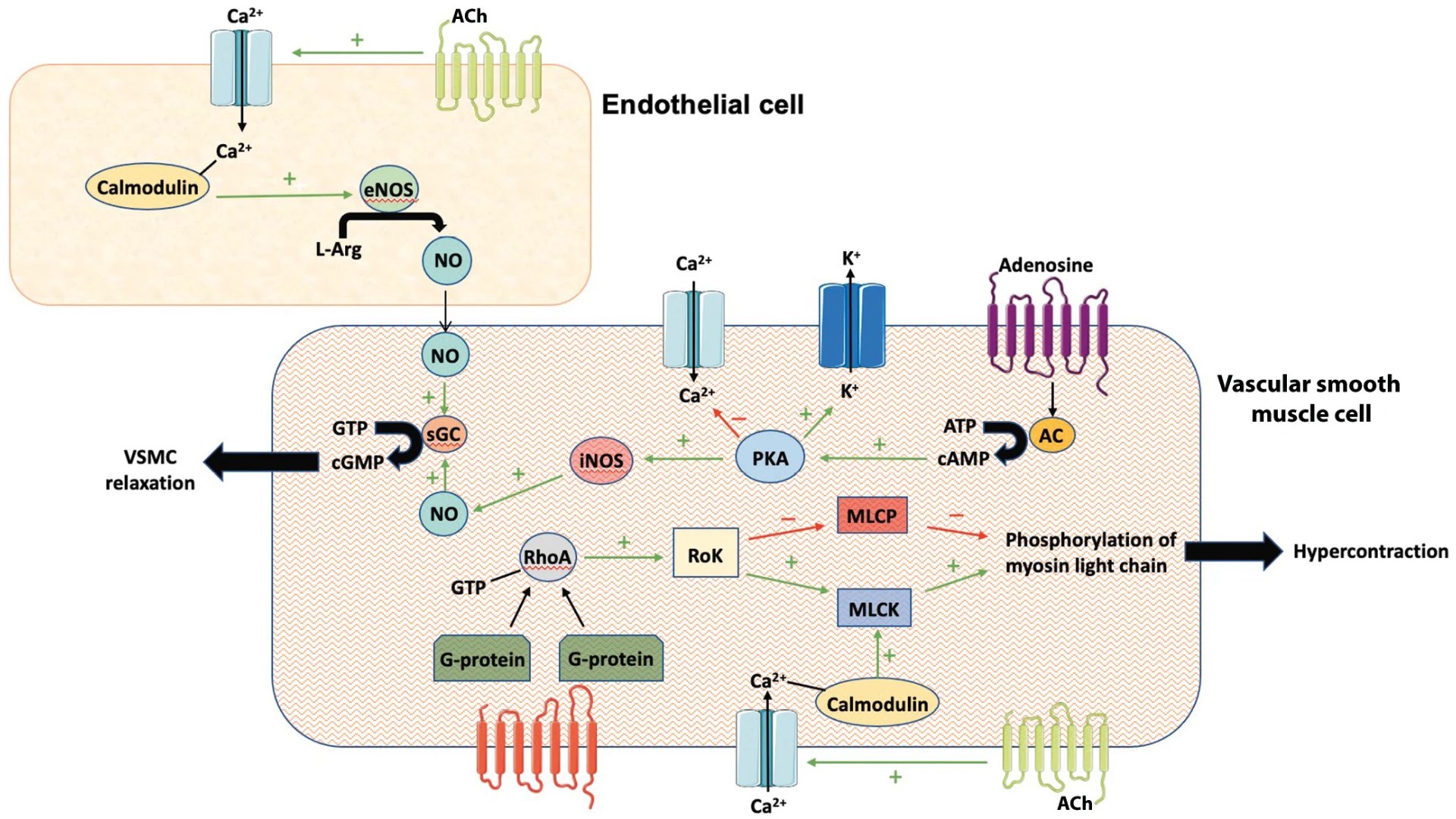
Figure 1. Cellular interplay between endothelial- and VSMC-mediated signalling pathways that promote relaxation and contraction. AC: adenylyl cyclase; ACh: acetylcholine; ATP: adenosine triphosphate; CA2+: calcium; cAMP: cyclic adenosine monophosphate; cGMP: cyclic guanosine monophosphate; eNOS: endothelial nitric oxide synthase; GTP: guanosine triphosphate; iNOS: inducible nitric oxide synthase; L-Arg: L-arginine; K+: potassium; MLCK: myosin light chain kinase; MLCP: myosin light chain phosphate; NO: nitric oxide; PKA: protein kinase A; RhoA: Ras homolog family member A; RoK: Rho-kinase; sGC: soluble guanylate cyclase; VSMC: vascular smooth muscle cell
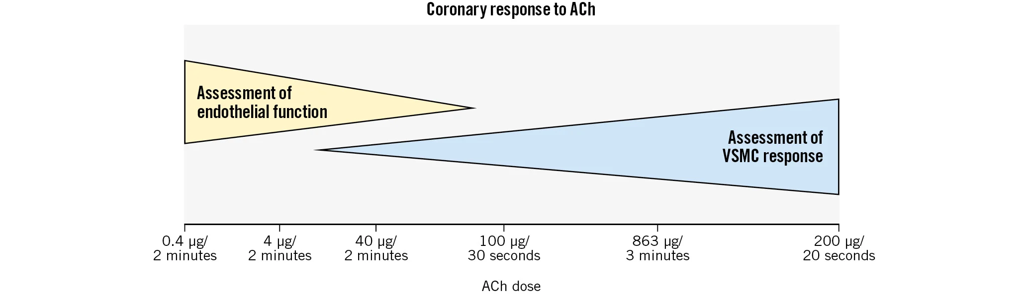
Figure 2. Acetylcholine (ACh) allows diagnosis of endothelium-dependent CMD and CS. Endothelial function is assessed at lower doses of ACh, while patients with CS have VSMC-mediated vasoconstriction at higher doses of ACh. The overlap between the two doses makes it difficult to reliably differentiate endothelium-dependent CMD from isolated microvascular spasm. The x-axis includes common dosing patterns used in different protocols worldwide. CMD: coronary microvascular dysfunction; CS: coronary spasm; VSMC: vascular smooth muscle cell


